What can we expect to find in a South American 17th-century table cabinet? Jewels? Letters from a long-distance lover? Makeup? Well, maybe. However, would we expect a skeleton? No, and neither did the scientific research and conservation staff of the Victoria and Albert Museum in London.
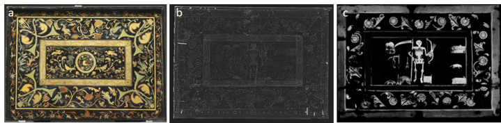
A recent paper by Burgio et al. details a technical study of a 17th-century table cabinet from the northern zone of the Vice-royalty of Peru (an area, which today encompasses parts of Peru, Colombia, and Ecuador). This study led to the first ever documented intentional use of calomel (mercury(I) chloride) as a white pigment in an art artifact, and to the first (more macabre) discovery of a hidden skeleton decoration with a scythe, a bow, and arrows (Figure 1).
The museum staff studied the South American table cabinet, which was a gift to the museum, with the initial intention of confirming the use of the barniz de Pasto technique. Barniz de Pasto is a type of Iberian American ‘lacquer,’ which involves heating and chewing the transparent resin, mopa mopa, (with or without pigments or dyes added) until it is pliable enough to be spread over and pressed onto a surface. The use of the barniz de Pasto technique was quickly confirmed using Fourier transform infrared spectroscopy (FTIR), which detected the presence of mopa mopa resin.
However, the use of further non-destructive and micro-destructive techniques led to more discoveries about the history of the cabinet. X-ray radiography (Victoria and Albert Museum, London) detected the presence of a heavy element which was able to block X-rays, but not on the surface of the cabinet. Micro-computed X-ray tomography (µCT, Natural History Museum London) was used to investigate this further and revealed a quite sinister design with a different fauna and flora pattern lying beneath the outermost surface of the inner lid (Figure 1). Macro X-ray fluorescence scanning (MA-XRF, National Gallery London) was then used to determine the elemental composition of the paint used in this hidden layer.
The white painted areas detected using X-ray radiography were found to contain mercury as the primary element using µXRF. Usually, the presence of mercury in artworks is associated with red, pink or orange due to the presence of vermilion (mercury(II) sulfide). However, the mercury-rich regions of the paint cross-sections were white with no shades of red, and X-ray diffraction (XRD) and Raman spectroscopy were used in conjunction to identify the pigment as calomel.
The skeleton layer (Figure 1c) had been covered at some point with a fauna and flora design (Figure 1a), making it invisible to the naked eye. But when was it covered? Again, scientific analyses can help us answer the question. XRF and Raman spectroscopy were used to identify titanium white (TiO2), commercially available in the 1920s, and phthalocyanine blue pigments, commercially available in 1935, on the outermost layers of the inner lid. These identifications push the terminus post quem (earliest possible date) to 1935.
The µCT results collected at the Natural History Museum London, which show the hidden layer of paint on the cabinet in great detail, are shown in the video below. This technique allowed researchers to virtually remove the outermost surface, or overpaint, of the inner lid.
Micro-CT (computed tomography) scan of the lid showing the hidden decorative scheme © the Trustees of the Natural History Museum
These results illustrate that appearances can be misleading. While walking around a museum, what you see might not be what was originally intended by the artist. However, developments in imaging techniques, complimentary characterization tools, and collaborations between different research labs have significantly improved our understanding of these “hidden artworks.” Now the only question left is whether or not it would be ethical to remove the 20th-century overpaint layer to reveal the original spooky design beneath it. But that’s for another time…
With Halloween around the corner, we highly recommend that you read more about the skeleton in the box!
All images adapted with permission from the Victoria and Albert Museum, London and the Natural History Museum, London.

4 thoughts on “Hidden Skeleton: Using scientific imaging tools to reveal the previous lives of art objects”